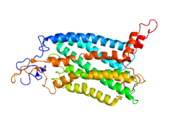
Découvert en 1994 [5], le CCR2 (C-C chemokine receptor type 2) est une protéine ayant une fonction de récepteur de la protéine CCL2. Il appartient à la famille des récepteurs aux chémokines CC. Son gène est le CCR2 situé sur le chromosome 3 humain. Chez l'homme, il existe deux isoformes CCR2A et CCR2B qui diffèrent par leur C-terminal, ce qui peut entraîner des propriétés de signalisation différentes[6].
L'activation du CCR2 par le CCL2 favorise largement la progression et les métastases d'une tumeur en attirant les monocytes suppressifs et les lymphocytes T régulateurs[21],[22], bien que comme toute liaison d'une chimiokine avec son récepteur peut avoir une action à la fois pro-tumorale et anti-tumorale[23].
Ils interviendraient dans la genèse de l'athérome[24]. L'inhibition de ce récepteur permettrait également de diminuer l'inflammation lors d'un infarctus du myocarde et d'améliorer son remodelage, du moins sur un modèle animal[25].
Sa stimulation favoriserait la migration de certaines cellules souches sanguines aux endroits où survient une inflammation[26].
Chez l'homme, l'inhibition de ce récepteur par un anticorps monoclonal diminue le taux sanguin de CRP[27].
Notes et références
↑ ab et cGRCh38: Ensembl release 89: ENSG00000121807 - Ensembl, May 2017
↑ ab et cGRCm38: Ensembl release 89: ENSMUSG00000049103 - Ensembl, May 2017
↑« Publications PubMed pour l'Homme », sur National Center for Biotechnology Information, U.S. National Library of Medicine
↑« Publications PubMed pour la Souris », sur National Center for Biotechnology Information, U.S. National Library of Medicine
↑(en) I F Charo, S J Myers, A Herman et C Franci, « Molecular cloning and functional expression of two monocyte chemoattractant protein 1 receptors reveals alternative splicing of the carboxyl-terminal tails. », Proceedings of the National Academy of Sciences, vol. 91, no 7, , p. 2752–2756 (ISSN0027-8424 et 1091-6490, PMID8146186, PMCIDPMC43448, DOI10.1073/pnas.91.7.2752, lire en ligne, consulté le )
↑Liyang Fei, Xiaochen Ren, Haijia Yu et Yifan Zhan, « Targeting the CCL2/CCR2 Axis in Cancer Immunotherapy: One Stone, Three Birds? », Frontiers in Immunology, vol. 12, (ISSN1664-3224, PMID34804061, PMCIDPMC8596464, DOI10.3389/fimmu.2021.771210, lire en ligne, consulté le )
↑Charo I, Myers S, Herman A, Fanci C, Connolly A, Coughlin S, Molecular cloning and functional expression of two monocyte chemoattractant protein 1 receptors reveals alternative splicing of the carboxyl-terminal tails, Proc Natl Acad Sci U S A, 1994;91:2752–2756
↑(en) Jee H. Lee, Seung G. Kang et Chang H. Kim, « FoxP3+ T Cells Undergo Conventional First Switch to Lymphoid Tissue Homing Receptors in Thymus but Accelerated Second Switch to Nonlymphoid Tissue Homing Receptors in Secondary Lymphoid Tissues », The Journal of Immunology, vol. 178, no 1, , p. 301–311 (ISSN0022-1767 et 1550-6606, DOI10.4049/jimmunol.178.1.301, lire en ligne, consulté le )
↑(en) Raffaella Bonecchi, Giancarlo Bianchi, Paola Panina Bordignon et Daniele D'Ambrosio, « Differential Expression of Chemokine Receptors and Chemotactic Responsiveness of Type 1 T Helper Cells (Th1s) and Th2s », The Journal of Experimental Medicine, vol. 187, no 1, , p. 129–134 (ISSN0022-1007 et 1540-9538, PMID9419219, PMCIDPMC2199181, DOI10.1084/jem.187.1.129, lire en ligne, consulté le )
↑Nansen A, Marker O, Bartholdy C, Thomsen AR. CCR2+ and CCR5+ CD8+ T Cells Increase During Viral Infection and Migrate to Sites of Infection. Eur J Immunol (2000) 30(7):1797–806. doi: 10.1002/1521-4141(200007)30:7<1797::AID-IMMU1797>3.0.CO;2-B
↑(en) Leonid S. Metelitsa, Hong-Wei Wu, Hong Wang et Yujun Yang, « Natural Killer T Cells Infiltrate Neuroblastomas Expressing the Chemokine CCL2 », The Journal of Experimental Medicine, vol. 199, no 9, , p. 1213–1221 (ISSN1540-9538 et 0022-1007, PMID15123743, PMCIDPMC2211904, DOI10.1084/jem.20031462, lire en ligne, consulté le )
↑(en) Duncan R. McKenzie, Ervin E. Kara, Cameron R. Bastow et Timona S. Tyllis, « IL-17-producing γδ T cells switch migratory patterns between resting and activated states », Nature Communications, vol. 8, no 1, (ISSN2041-1723, PMID28580944, PMCIDPMC5465362, DOI10.1038/ncomms15632, lire en ligne, consulté le )
↑Frade J, Mellado M, del Real G, Gutierrez-Ramos J, Lind P, Martinez-A C. Characterization of the CCR2 Chemokine Receptor: Functional CCR2 Receptor Expression in B Cells. J Immunol (1997) 159(11):5576–84.
↑(en) M. Swiecki, H.L. Miller, R. Sesti-Costa et M. Cella, « Microbiota induces tonic CCL2 systemic levels that control pDC trafficking in steady state », Mucosal Immunology, vol. 10, no 4, , p. 936–945 (PMID27827374, PMCIDPMC5423869, DOI10.1038/mi.2016.99, lire en ligne, consulté le )
↑(en) Qingjun Pan, Yongmin Feng, Yanxia Peng et Hongjiu Zhou, « Basophil Recruitment to Skin Lesions of Patients with Systemic Lupus Erythematosus Mediated by CCR1 and CCR2 », Cellular Physiology and Biochemistry, vol. 43, no 2, , p. 832–839 (ISSN1015-8987 et 1421-9778, DOI10.1159/000481609, lire en ligne, consulté le )
↑(en) Fikru Belema-Bedada, Shizuka Uchida, Alessandra Martire et Sawa Kostin, « Efficient Homing of Multipotent Adult Mesenchymal Stem Cells Depends on FROUNT-Mediated Clustering of CCR2 », Cell Stem Cell, vol. 2, no 6, , p. 566–575 (DOI10.1016/j.stem.2008.03.003, lire en ligne, consulté le )
↑(en) Kim S. C. Weber, Peter J. Nelson, Hermann-Joseph Gröne et Christian Weber, « Expression of CCR2 by Endothelial Cells: Implications for MCP-1 Mediated Wound Injury Repair and In Vivo Inflammatory Activation of Endothelium », Arteriosclerosis, Thrombosis, and Vascular Biology, vol. 19, no 9, , p. 2085–2093 (ISSN1079-5642 et 1524-4636, DOI10.1161/01.ATV.19.9.2085, lire en ligne, consulté le )
↑(en) Joseph El Khoury, Michelle Toft, Suzanne E Hickman et Terry K Means, « Ccr2 deficiency impairs microglial accumulation and accelerates progression of Alzheimer-like disease », Nature Medicine, vol. 13, no 4, , p. 432–438 (ISSN1078-8956 et 1546-170X, DOI10.1038/nm1555, lire en ligne, consulté le )
↑(en) Gordon L. Warren, Tracy Hulderman, Dawn Mishra et Xin Gao, « Chemokine receptor CCR2 involvement in skeletal muscle regeneration », The FASEB Journal, vol. 19, no 3, , p. 1–23 (ISSN0892-6638 et 1530-6860, DOI10.1096/fj.04-2421fje, lire en ligne, consulté le )
↑(en) Qiongyu Hao, Jaydutt V. Vadgama et Piwen Wang, « CCL2/CCR2 signaling in cancer pathogenesis », Cell Communication and Signaling, vol. 18, no 1, (ISSN1478-811X, PMID32471499, PMCIDPMC7257158, DOI10.1186/s12964-020-00589-8, lire en ligne, consulté le )
↑(en) Bin-Zhi Qian, Jiufeng Li, Hui Zhang et Takanori Kitamura, « CCL2 recruits inflammatory monocytes to facilitate breast-tumour metastasis », Nature, vol. 475, no 7355, , p. 222–225 (ISSN0028-0836 et 1476-4687, PMID21654748, PMCIDPMC3208506, DOI10.1038/nature10138, lire en ligne, consulté le )
↑(en) Pierre-Louis Loyher, Juliette Rochefort, Camille Baudesson de Chanville et Pauline Hamon, « CCR2 Influences T Regulatory Cell Migration to Tumors and Serves as a Biomarker of Cyclophosphamide Sensitivity », Cancer Research, vol. 76, no 22, , p. 6483–6494 (ISSN0008-5472 et 1538-7445, DOI10.1158/0008-5472.CAN-16-0984, lire en ligne, consulté le )
↑(en) Aleksandra J. Ozga, Melvyn T. Chow et Andrew D. Luster, « Chemokines and the immune response to cancer », Immunity, vol. 54, no 5, , p. 859–874 (PMID33838745, PMCIDPMC8434759, DOI10.1016/j.immuni.2021.01.012, lire en ligne, consulté le )
↑Boring L, Gosling J, Cleary M, Charo IF, Decreased lesion formation in CCR2-/- mice reveals a role for chemokines in the initiation of atherosclerosis, Nature, 1998;394:894–897
↑Majmudar M, Keliher E, Heidt T et al. Monocyte-directed RNAi targeting CCR2 improves infarct healing in atherosclerosis-prone mice, Circulation, 2013;127:2038–2046
↑Si Y, Tsou CL, Croft K, Charo IF, CCR2 mediates hematopoietic stem and progenitor cell trafficking to sites of inflammation in mice, J Clin Invest, 2010;120:1192–1203
↑Gilbert J, Lekstrom-Himes J, Donaldson D et al. Effect of CC chemokine receptor 2 CCR2 blockade on serum C-reactive protein in individuals at atherosclerotic risk and with a single nucleotide polymorphism of the monocyte chemoattractant protein-1 promoter region, Am J Cardiol, 2011;107:906–911


 Portail de la médecine
  Portail de la biologie cellulaire et moléculaire


